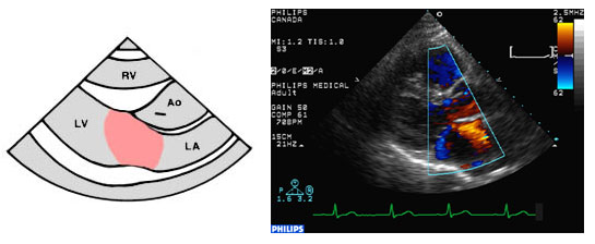
Normal blood flow is usually laminar, characterized by a smooth homogeneous color pattern. Aliasing will occur if the velocities exceed the Nyquist limit. You can image normal blood flow from any window that lets you see both proximal and distal to a valve.
You can see blood flow through the mitral valve (MV) from the parasternal, apical and subcostal views. Normal left ventricular inflow occurs in diastole, as blood moves from the left atrium to the left ventricle.
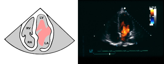
This apical four-chamber view shows normal diastolic blood flow through the MV. An enhanced velocity map has primarily red colors due to the flow toward the transducer. Oranges and yellows indicate the higher velocities in the center of the jet. The smooth progression from red to yellow indicates laminar blood flow.
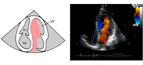
Though it looks significantly different from the picture above, the diastolic left ventricular inflow depicted in this apical four-chamber view is normal. This image also has an enhanced velocity map. The smooth progresion from red to blue indicates laminar blood flow.