32
(2005-04-20 15:57:07) 评论 (0)| Blood Flow Patterns in the Heart: Blood Flow Across Stenotic Sites | |||||||||||||||||||||
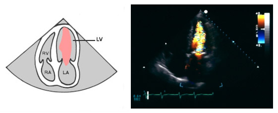
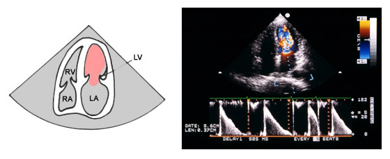
Blood flow across stenotic sites is often high velocity, and generally aliases. As the velocity increases, the likelihood of turbulence also increases. To visualize blood flow across a stenotic site, use any view that shows both the obstruction and the chamber distal to it. To estimate the severity of a stenotic lesion calculate the pressure gradient across the obstruction. CFI identifies blood flow characteristics and jet direction, but does not produce quantifiable numbers. The colors indicate only relative mean velocity estimates, so they are not useful in determining severity. However, you can use the blood flow spatial information CFI provides to quickly guide Doppler beam placement.
| |||||||||||||||||||||


